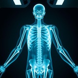
1. Right kidney (red)
2. Left kidney (red)
3. Location of right ureter (white outline) – not normally visualized
4. Location of left ureter (white outline) – not normally visualized
5. Urinary bladder (orange)
6. Gas in the rectum (green)
7. Location of right adrenal gland (white outline) – not normally visualized
8. Location of left adrenal gland (white outline) – not normally visualized
9. Location of the gallbladder (white outline) – not normally visualized
https://t.me/radio_2023
2. Left kidney (red)
3. Location of right ureter (white outline) – not normally visualized
4. Location of left ureter (white outline) – not normally visualized
5. Urinary bladder (orange)
6. Gas in the rectum (green)
7. Location of right adrenal gland (white outline) – not normally visualized
8. Location of left adrenal gland (white outline) – not normally visualized
9. Location of the gallbladder (white outline) – not normally visualized
https://t.me/radio_2023
❤8👍5💯1
1. Liver (purple)
2. Spleen (pink)
3. Location of the pancreas (white outline) – not normally visualized
https://t.me/radio_2023
2. Spleen (pink)
3. Location of the pancreas (white outline) – not normally visualized
https://t.me/radio_2023
❤2👍2❤🔥1
✅ pneumoperitoneum
👉 Characteristics
- Free intra-abdominal air.
- May follow surgery. It is normal to see free air up to 3 days following a laparotomy. Beyond 3 days, there is a high suspicion of air leak from an
abdominal/pelvic viscus.
- Associated with a perforated abdominal or pelvic viscus (e.g. duodenal ulcer, ruptured appendicitis or diverticulitis, vaginal injury), blunt or penetrating trauma, necrotising enterocolitis, inflammatory bowel disease.
- Pneumomediastinum may track into the abdomen.
- Rupture of a bleb from pneumatosis cystoides intestinalis may lead to
asymptomatic intraperitoneal air.
👉 Clinical Features
🚫 - THIS IS A SURGICAL EMERGENCY.
- Abdominal pain. Vomiting. Diarrhoea.
- Pyrexia. Sweating.
- Patient shock – tachycardia and hypotension.
- Abdominal tenderness with signs of peritonism (guarding and rebound tenderness).
👉 Radiological Features
- As little as 5 ml of air may be visible.
📌- Erect CXR – free air lying under one or both of the hemidiaphragms.
- The CXR may be normal or if supine/semi-recumbent, it may be missed.
📌 - AXR or decubitus films may be helpful
- Locules of gas lying outside bowel often with odd/linear margins.
- Both sides of the bowel may be visible (Rigler’s sign).
- The falciform ligament may be demonstrated.
- Hyperlucency overlying the liver with clear inferior hepatic margin outlined by air.
📌 - CT – demonstrates even tiny locules of free air if clinically suspicious. Also allows assessment of underlying cause.
https://t.me/radio_2023
👉 Characteristics
- Free intra-abdominal air.
- May follow surgery. It is normal to see free air up to 3 days following a laparotomy. Beyond 3 days, there is a high suspicion of air leak from an
abdominal/pelvic viscus.
- Associated with a perforated abdominal or pelvic viscus (e.g. duodenal ulcer, ruptured appendicitis or diverticulitis, vaginal injury), blunt or penetrating trauma, necrotising enterocolitis, inflammatory bowel disease.
- Pneumomediastinum may track into the abdomen.
- Rupture of a bleb from pneumatosis cystoides intestinalis may lead to
asymptomatic intraperitoneal air.
👉 Clinical Features
🚫 - THIS IS A SURGICAL EMERGENCY.
- Abdominal pain. Vomiting. Diarrhoea.
- Pyrexia. Sweating.
- Patient shock – tachycardia and hypotension.
- Abdominal tenderness with signs of peritonism (guarding and rebound tenderness).
👉 Radiological Features
- As little as 5 ml of air may be visible.
📌- Erect CXR – free air lying under one or both of the hemidiaphragms.
- The CXR may be normal or if supine/semi-recumbent, it may be missed.
📌 - AXR or decubitus films may be helpful
- Locules of gas lying outside bowel often with odd/linear margins.
- Both sides of the bowel may be visible (Rigler’s sign).
- The falciform ligament may be demonstrated.
- Hyperlucency overlying the liver with clear inferior hepatic margin outlined by air.
📌 - CT – demonstrates even tiny locules of free air if clinically suspicious. Also allows assessment of underlying cause.
https://t.me/radio_2023
Telegram
basic radiology
X-ray Chanel
https://www.facebook.com/profile.php?id=61558872729627&mibextid=ZbWKwL
https://www.facebook.com/profile.php?id=61558872729627&mibextid=ZbWKwL
❤6👍2
Forwarded from basic radiology
Radiological modalities used
in hepatobiliary system
🔻 X-Ray
✏️ Advantages.
▪️ Cheap.
▪️ Widely available
✏️ Disadvantages.
▪️ Radiation.
▪️ Poor soft tissue details.
🔷 COMPUTED TOMOGRAPHY (CT)
🔻 Advantages.
▪️ Good at evaluating abdominal solid organs.
🔸 Disadvantages
▪️ Contrast can be contraindicated.
🟪 MRI
🔹 Advantages.
● Good at evaluating solid organs.
● No radiation
🔸 Disadvantages
● Long scan time.
● Less available than other modalities
🟥 ULTRASOUND
🟡 Advantages.
● No radiation.
● Very good soft tissue details.
🟢 Disadvantages.
● Operator dependant.
● Limited in evaluating bowel and calcification.
🟥 NUCLEAR MEDICINE.
🔷 Advantages.
▪️Excellent in evaluating organ function.
🟫 Disadvantages.
▪️ Radiation.
▪️Very poor in evaluating anatomy.
▪️ Not widely available.
in hepatobiliary system
🔻 X-Ray
✏️ Advantages.
▪️ Cheap.
▪️ Widely available
✏️ Disadvantages.
▪️ Radiation.
▪️ Poor soft tissue details.
🔷 COMPUTED TOMOGRAPHY (CT)
🔻 Advantages.
▪️ Good at evaluating abdominal solid organs.
🔸 Disadvantages
▪️ Contrast can be contraindicated.
🟪 MRI
🔹 Advantages.
● Good at evaluating solid organs.
● No radiation
🔸 Disadvantages
● Long scan time.
● Less available than other modalities
🟥 ULTRASOUND
🟡 Advantages.
● No radiation.
● Very good soft tissue details.
🟢 Disadvantages.
● Operator dependant.
● Limited in evaluating bowel and calcification.
🟥 NUCLEAR MEDICINE.
🔷 Advantages.
▪️Excellent in evaluating organ function.
🟫 Disadvantages.
▪️ Radiation.
▪️Very poor in evaluating anatomy.
▪️ Not widely available.
❤7👍1
Forwarded from basic radiology
Sammary of common CT signs in pulmonary diseases
CT Lung Signs in Pulmonary Diseases - One Page Summary of Pulmonary Signs
- Air Crescent sign
- Monod sign
- Halo sign
- Atoll sign (reverse-halo sign)
- Cheerio sign
- Comet tail sign
- Corona radiata (sunburst sign)
- Crazy paving sign
- Galaxy sign
- Mosaic attenuation
- Head cheese sign
- Sant storm sign
- Water lily sign (Camalote sign)
- Air bronchogram sign
- Bronchus sign
- Signet ring sign
- Tram track sign
- Tree-in-bud sign
- CT angiogram sign
- Feeding vessel sign
- Polo mint sign
- Split pleura sign
CT Lung Signs in Pulmonary Diseases - One Page Summary of Pulmonary Signs
- Air Crescent sign
- Monod sign
- Halo sign
- Atoll sign (reverse-halo sign)
- Cheerio sign
- Comet tail sign
- Corona radiata (sunburst sign)
- Crazy paving sign
- Galaxy sign
- Mosaic attenuation
- Head cheese sign
- Sant storm sign
- Water lily sign (Camalote sign)
- Air bronchogram sign
- Bronchus sign
- Signet ring sign
- Tram track sign
- Tree-in-bud sign
- CT angiogram sign
- Feeding vessel sign
- Polo mint sign
- Split pleura sign
❤4❤🔥1👍1
② Monad's sign.
The axial CT scan of the chest, on the left, shows a nodule (black arrow) within an air-filled cavity with a crescent of air at the superior surface of the cavity (white arrow). In the close-up view of the left upper lobe from a chest radiograph of another person, on the right, there is again seen a thin-walled cavity (white arrow) containing a soft-tissue mass (black arrow). Both of these cavities were from old tuberculosis.
▊ Imaging Findings
In secondary, non-invasive (saprophytic) aspergillosis
The fungus ball is usually freely movable and will move to the dependent surface on decubitus radiographs or supine and prone CT studies
Rarely, the fungus ball may calcify
Invasive aspergillosis
On CT, there may be ground glass densities in the area around the nodular density called the "halo sign" and indicative of hemorrhage
● Differential Diagnosis
Cavitating neoplasm
TB
Nocardiosis
The axial CT scan of the chest, on the left, shows a nodule (black arrow) within an air-filled cavity with a crescent of air at the superior surface of the cavity (white arrow). In the close-up view of the left upper lobe from a chest radiograph of another person, on the right, there is again seen a thin-walled cavity (white arrow) containing a soft-tissue mass (black arrow). Both of these cavities were from old tuberculosis.
▊ Imaging Findings
In secondary, non-invasive (saprophytic) aspergillosis
The fungus ball is usually freely movable and will move to the dependent surface on decubitus radiographs or supine and prone CT studies
Rarely, the fungus ball may calcify
Invasive aspergillosis
On CT, there may be ground glass densities in the area around the nodular density called the "halo sign" and indicative of hemorrhage
● Differential Diagnosis
Cavitating neoplasm
TB
Nocardiosis
❤4👍2❤🔥1💯1
➡️ Halo sign: A pulmonary nodule or mass is surrounded by Ground Glass appearance.
➡️ Reversed Halo Sign: Focal Rounded area of Ground Glass opacity surrounded by a complete ring of consolidation.
➡️ Reversed Halo Sign: Focal Rounded area of Ground Glass opacity surrounded by a complete ring of consolidation.
❤4👍1💯1
③ Cheerio sign in thoracic 🩻
relates to pulmonary nodules with a central lucent cavity supplied by a patent bronchus as seen on CT.
Due to proliferation of (malignant or non-malignant) cells around an airway.
May be found in Pt's w Langerhans Cell H. or 🫁AdenoCa.
https://t.me/radio_2023
relates to pulmonary nodules with a central lucent cavity supplied by a patent bronchus as seen on CT.
Due to proliferation of (malignant or non-malignant) cells around an airway.
May be found in Pt's w Langerhans Cell H. or 🫁AdenoCa.
https://t.me/radio_2023
❤7👍5💯2