Mesoblastic Nephroma
🌀Solid tumor of infancy, born with it
🌀Antenatal USG: ~Polyhydroamnios
🌀No invasion of;
➖Collecting system
➖Venous system
🌀Often involve renal sinus
MZeba
🌀Solid tumor of infancy, born with it
🌀Antenatal USG: ~Polyhydroamnios
🌀No invasion of;
➖Collecting system
➖Venous system
🌀Often involve renal sinus
MZeba
👍2
NAI#
🦴 LL#in non ambulant👶
🦴 Metaphyseal corner/🪣 handle#
🦴Multiple#of Different age
🦴Scapula#
🦴Post rib#
🦴Sternal#
🦴Non parietal💀/#crossing suture
MZeba
🦴 LL#in non ambulant👶
🦴 Metaphyseal corner/🪣 handle#
🦴Multiple#of Different age
🦴Scapula#
🦴Post rib#
🦴Sternal#
🦴Non parietal💀/#crossing suture
MZeba
👍2
IMAGING IN PEDIATRICS
Day1⃣ : Pedia MSK
📚Core(old): page 802-824
📚Crack: Section 13,14
🎥 https://www.youtube.com/playlist?list=PLsYxTl3AgoMP02IMOdHsmuhoBdR90vOJG
Day2⃣ : Pedia GUT
📚Core(old): 791-801
📚Crack: Section 9,10,11&12
🎥 https://www.youtube.com/playlist?list=PLsYxTl3AgoMMOE3DStypi7Jj_Cb8pHjrO
Day3⃣: Pedia GIT
📚Core(old): 770-790
📚Crack: Section 7,8
🎥 https://www.youtube.com/playlist?list=PLsYxTl3AgoMPoNytJrmfegJBW80H1reIV
Day4⃣: Pedia Airway,💙& Chest
📚Core(old): page 742-769
📚Crack: Section 4,5,6
🎥 https://www.youtube.com/playlist?list=PLsYxTl3AgoMN4Vkfr4K7AXoFqc4IbTpB3
Day5⃣: Pedia CNS
📚Core(old):825-835
📚Crack: Section 1,2 & 3
🎥 https://www.youtube.com/playlist?list=PLsYxTl3AgoMP_rrHW10XCQoBYOHCRKaQ2
MZeba
👍1
#FRCR2B2024
Teaching files / Case Reviews
1️⃣ MZeba Playlist https://youtube.com/playlist?list=PLsYxTl3AgoMP0Zf3bmnhfSH-F8ABOGr8t
2️⃣ https://youtube.com/playlist?list=PLYcjsMRiuBVBcpJHZBSyKxbxhmS6Deugz
3️⃣
4️⃣
5️⃣
Teaching files / Case Reviews
1️⃣ MZeba Playlist https://youtube.com/playlist?list=PLsYxTl3AgoMP0Zf3bmnhfSH-F8ABOGr8t
2️⃣ https://youtube.com/playlist?list=PLYcjsMRiuBVBcpJHZBSyKxbxhmS6Deugz
3️⃣
4️⃣
5️⃣
👍1
Forwarded from FRCR 2B 2023/2024
TF#24
Pleuropulmonary Blastoma
💎Big chest mass 1-2Y, R>L, Pleural based
💎Cystic type B9 younger kid
💎Solid type older kid, met to 🧠🦴
💎No calcification, no rib erosion
💎 10% associated with MLCN
MZeba
Pleuropulmonary Blastoma
💎Big chest mass 1-2Y, R>L, Pleural based
💎Cystic type B9 younger kid
💎Solid type older kid, met to 🧠🦴
💎No calcification, no rib erosion
💎 10% associated with MLCN
MZeba
👍5
OI
🔎Total lucent skull 💀
🔎Multiple fractures with hyperplastic callus
🔎Fibula longer than Tibia
🔎Wormian 🦴
🔎Blue sclera
🔎Otosclerosis
CTC#132
MZeba
🔎Total lucent skull 💀
🔎Multiple fractures with hyperplastic callus
🔎Fibula longer than Tibia
🔎Wormian 🦴
🔎Blue sclera
🔎Otosclerosis
CTC#132
MZeba
👍1
*Replogle tube*
🏎️ Used in esophageal atresia
🏎️ A double lumen tube which [in contrast to an NGT] has a number of side holes.
🏎️ The Replogle tube rests in the proximal oesophageal pouch to simultaneously irrigate and aspirate secretions/debris.
MZeba
🏎️ Used in esophageal atresia
🏎️ A double lumen tube which [in contrast to an NGT] has a number of side holes.
🏎️ The Replogle tube rests in the proximal oesophageal pouch to simultaneously irrigate and aspirate secretions/debris.
MZeba
👍9
Caffey Disease
💎Self limiting within 6M of life
💎Classic:Hot mandible on bone scan
💎Mandible,clavicle &Ulna
💎Fever, irritability, periosteal rxn
💎Coarse,irregular,asymmetric periosteal rxn➕soft tissue swelling over affected areas.
MZeba
💎Self limiting within 6M of life
💎Classic:Hot mandible on bone scan
💎Mandible,clavicle &Ulna
💎Fever, irritability, periosteal rxn
💎Coarse,irregular,asymmetric periosteal rxn➕soft tissue swelling over affected areas.
MZeba
👍3
ARPCKD Associations
1. Caroli disease
2. Congenital hepatic fibrosis: the degree of which is inversely proportional to the age of presentation
3. Multiple biliary hamartomas
(Radiopedia)
1. Caroli disease
2. Congenital hepatic fibrosis: the degree of which is inversely proportional to the age of presentation
3. Multiple biliary hamartomas
(Radiopedia)
👍5
Peads👦 SBA Quizzes by MZeba
--- MSK 🦴
1. http://t.me/QuizBot?start=7ZCBYEft
2. http://t.me/QuizBot?start=NYWP8qEz
3. http://t.me/QuizBot?start=7GHDWxhm
--- 🧠 CNS
1. http://t.me/QuizBot?start=aPOzhCrg
2. http://t.me/QuizBot?start=k3VsR5Vu
--- Chest 🫁
1.http://t.me/QuizBot?start=XdNqMmNm
2. http://t.me/QuizBot?start=tlGvggZt
--- 🤎CVS
1. http://t.me/QuizBot?start=tbwP2LML
2. http://t.me/QuizBot?start=7ZVAZuNd
--- GUT
1. http://t.me/QuizBot?start=Uk90C8cD
2. http://t.me/QuizBot?start=A2gLrS7d
--- GIT
1. http://t.me/QuizBot?start=kpfTtTfm
2. http://t.me/QuizBot?start=HFsVZRYj
--- Review Quizzes
1. http://t.me/QuizBot?start=4goMzkX5
2. http://t.me/QuizBot?start=LFFk9y5D
3. http://t.me/QuizBot?start=Y0ot6vMZ
4.http://t.me/QuizBot?start=jJcxPV35
5. http://t.me/QuizBot?start=S7UsxxzP
--- Challenging Questions from 📚
1. Lindsay: http://t.me/QuizBot?start=GVGYcxZ6
2.Currie: http://t.me/QuizBot?start=iW8GneNN
MZeba
Quiz Directory
SBAs on Imaging Skeletal Congenital Anomalies1⃣ by MZeba
Source: Currently available SBAs books / 20 questions
👍1
PaedsImagingQuizzesMZeba pinned «Peads👦 SBA Quizzes by MZeba --- MSK 🦴 1. http://t.me/QuizBot?start=7ZCBYEft 2. http://t.me/QuizBot?start=NYWP8qEz 3. http://t.me/QuizBot?start=7GHDWxhm --- 🧠 CNS 1. http://t.me/QuizBot?start=aPOzhCrg 2. http://t.me/QuizBot?start=k3VsR5Vu --- Chest 🫁 …»
HPS
⚀ Idiopathic, hypertrophy &hyperplasia= circular muscle pylorus
⚁ 1st borns, 2-12wk, projectile vomiting, olive shape mass ♂>♀
⚂ XR: 🐛sign
⚃ USG; ++gastric peristalsis, pyloric canal fail to open,
Pyloric canal leng➡14mm,
Pyloric canal diameter➡11mm
Muscle wall thick➡4mm
⚄ Paradoxical aciduria.
MZeba
🥧 π: 3.1415
👍1
Imaging of Nephroblastomatosis:
🌀Nephrogenic Rests: May precede Wilms tumor.
🌀Age Factor: Rare after 7 years old.
🌀Symptoms: Usually asymptomatic or presents as a flank mass.
🌀Associated Syndromes:
🔸Hemihypertrophy,
🔸Beckwith-Wiedemann syndrome,
🔸trisomy 18,
🔸sporadic aniridia.
🌀Progression: Most regress spontaneously; up to 33% may develop Wilms tumor.
🌀Screening: Ultrasound (US) every 3 months until age 7.
🌀US Appearance: Hypoechoic or isoechoic to renal parenchyma.
🌀CT Appearance: Homogeneous, low attenuation; enhances less than normal renal tissue.
MZeba
👍3
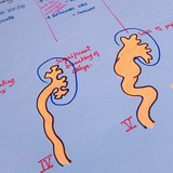
Wormian Bones Causes:
P: yknodysostosis
O: osteogenesis imperfecta
R: rickets
K: kinky hair syndrome
C: cleidocranial dysostosis
H: hypothyroidism/hypophosphatasia
O: otopalatodigital syndrome
P: primary acroosteolysis (Hajdu-Cheney)/pachydermoperiostosis/progeria
S: syndrome of Downs
(Radiopedia)
MZeba
P: yknodysostosis
O: osteogenesis imperfecta
R: rickets
K: kinky hair syndrome
C: cleidocranial dysostosis
H: hypothyroidism/hypophosphatasia
O: otopalatodigital syndrome
P: primary acroosteolysis (Hajdu-Cheney)/pachydermoperiostosis/progeria
S: syndrome of Downs
(Radiopedia)
MZeba
👍4
Multicystic Dysplastic Kidney (MCDK)
🌀Etiology: Often due to ureteral atresia or ureteropelvic junction obstruction in utero.
🌀Approximately 50% of cases show complete involution by age 7.
🌀Treatment: Resection is only necessary for complicated cases.
🌀Contralateral Kidney: Always check to rule out pathology; ~50% of cases have contralateral abnormalities: VUJ obstruction, VU reflux.
Imaging Findings:
🌀USG:
➖Multiple non-communicating cysts of varying sizes with no intervening renal parenchyma.
➖Normal renal cortex is not visualized.
🌀MAG 3
➖No uptake or excretion in the affected kidney.
🌀DD:
➖Distinguish from multilocular cystic nephroma, which typically does not involve the entire kidney
➖Hydronephrosis: Communicating cysts with a larger cyst in the center.
MZeba
👍3
NEC
Stage I
🌀Intestinal dilatation
Treatment
.. Oral feeding cessation
.. Parenteral nutrition
.. NGT suction
.. Antibiotics
Stage II
🌀 Intestinal dilatation
🌀PV gas
🌀 Pneumatosis intestinalis
Treatment
➕
Correction of metabolic acidosis
Stage IIIa:
🌀Shock
🌀Ascites
Treatment
Same as in stage II
Stage IIIb
🌀 Perforation
🌀 Pneumoperitoneum
Treatment: Surgery
(Radiopedia)
MZeba
👍1
Midgut Volvulus:
🌀Radiologic Emergency.
🌀Associated Conditions:
➖Congenital diaphragmatic hernia,
➖gastroschisis
➖omphalocele.
🌀Cecum Position: Malrotation often involves an abnormal cecal position, but a normal enema does not rule out malrotation.
🌀Cross-Sectional Imaging:
➖Abnormal SMA and SMV relationship; SMV should be anterior and to the right of SMA.
➖Whirlpool sign of twisted mesentery
🌀On UGI study:
➖corkscrew sign
➖tapering or beaking of the bowel in complete obstruction
➖malrotated bowel configuration
MZeba
👍3
Choanal Atresia:
🌀Two types: Bony (90%) and Membraneous (10%)
🌀It’s usually unilateral (65%)
🌀There is a known association with early
pregnancy use of anti thyroid drugs.
🌀There are multiple syndromes associated with this - the big one to know is CHARGE (Coloboma.
Heart Defects, Atresia - Choanal, Retardation.
Genital Issues, Ear Problems
(Source: CTC-CC)
👍3
CPAM
🌀Best Imaging Modalities
➖Prenatal: USG (first-line) – Cystic/hyperechoic lung lesion, MRI for better differentiation.
➖ Postnatal: Chest X-ray (cystic/solid lung mass), CT chest (Gold Standard) – Defines cystic vs. solid components, surgical planning.
🌀CPAM Types (Stocker Classification)
➖Type 1 (Most common, 70%) – Large air-filled cysts (>2 cm).
➖Type 2 (20%) – Small cysts (<2 cm), mixed solid-cystic.
➖Type 3 (10%) – Solid, microcystic mass-like.
➖Type 4 (Rare) – Large cystic, mimics pneumothorax.
🌀Differentiation on Imaging
➖CPAM vs. BPS: CPAM lacks systemic arterial supply (confirm with CT angiography).
➖CPAM vs. CDH: CPAM has no bowel loops in thorax (unlike diaphragmatic hernia).
🌀Complications
➖Infection: Air-fluid levels in cysts.
➖Malignancy Risk: Rare (e.g., pleuropulmonary blastoma).
➖Hydrops Fetalis: Seen in large prenatal lesions.
🌀Management
➖Small/asymptomatic: Follow-up imaging.
➖Large/symptomatic: Surgical resection to prevent complications. MZeba
👍10