This media is not supported in your browser
VIEW IN TELEGRAM
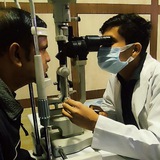
Retinal Tear - Retinal Detachment 👁
👍5❤3
Figure 1. Illustration depicting the zones, stages, and types of ROP. Zone I encompasses the circular area centered on the optic nerve head, having a radius equal to twice the distance between the optic nerve and the fovea, while Zone II extends as a circle centered on the optic nerve head, presenting a radius equal to the distance between the optic nerve and nasal ora serrata. Zone III covers the peripherical retinal area, extending over Zone II. Stage 1 manifests as a demarcation line, delineating the boundary between the physiologically vascularized retina and the peripheral avascular retina. In Stage 2, this line progresses into a distinct ridge. Stage 3 marks the onset of extraretinal neovascularization and hemorrhages. Stage 4 indicates partial and stage 5 total retinal detachment, respectively. The plus disease is characterized by pronounced vascular dilation and tortuosity.
Type 1: high-risk pre-threshold ROP includes Zone 1 with + disease at any stage, Zone 1 stage 3 without + disease, or Zone 2 stage 2 or 3 with + disease, necessitating prompt therapy.
Type 2: low-risk pre-threshold ROP comprises Zone 1 stage 1 or stage 2 without + disease, and Zone 2 stage 3 without + disease, recommended for follow-up.
👍4❤2🔥1
Please open Telegram to view this post
VIEW IN TELEGRAM
Identify the syndrome👆
❤1
Please open Telegram to view this post
VIEW IN TELEGRAM
DraftOpthalmicAssistant05June2026.pdf
3.6 MB
DraftOpthalmicAssistant05June2026.pdf