Forwarded from 0/0 (Haidar A. Fahad)
The arrow points to a perivalvular abcess near the aortic valve due to IE (Infective Endocarditis)
Forwarded from 0/0 (Haidar A. Fahad)
When your P and QRS waves are regular, but are dissociated and do not sync together (كلٌّ يغني على ليلاه) this is 3rd degree AV block.
Forwarded from 0/0 (Haidar A. Fahad)
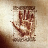
In the early 20th century, the physician was equipped primarily with a keen sense of observation and a compassionate heart; effective medications, and diagnostic laboratory tests; reliable imaging techniques were still to come. During house calls, the physician used his observational skills to evaluate both the surroundings and family members with respect to their limitations and benefits in regard to the patient. The observational skills of vision, hearing, touch, smell, and taste were well developed in most doctors. These skills were honed to razor-sharpness in the “diagnostician,” a term of honor applied to any physician, specialist, or nonspecialist, who was able to decipher complex clinical problems.
With the development of computed tomography (CT) and magnetic resonance imaging (MRI), as well as more sensitive and reliable laboratory tests in the late 1970s, the learned observational skills of physicians began to decline from disuse as reliance on these modalities for establishing diagnoses increased. Clinical medical decision making became unduly influenced by the tyranny of the tests, even though for many medical conditions—endocrine, infectious, malignant, and rheumatic, among others—the use of the eye and ear was sufficient to make the correct diagnosis or arrive at a limited group of diagnoses more rapidly and at much less cost.
With the development of computed tomography (CT) and magnetic resonance imaging (MRI), as well as more sensitive and reliable laboratory tests in the late 1970s, the learned observational skills of physicians began to decline from disuse as reliance on these modalities for establishing diagnoses increased. Clinical medical decision making became unduly influenced by the tyranny of the tests, even though for many medical conditions—endocrine, infectious, malignant, and rheumatic, among others—the use of the eye and ear was sufficient to make the correct diagnosis or arrive at a limited group of diagnoses more rapidly and at much less cost.
Forwarded from 0/0 (Haidar A. Fahad)
0/0
In the early 20th century, the physician was equipped primarily with a keen sense of observation and a compassionate heart; effective medications, and diagnostic laboratory tests; reliable imaging techniques were still to come. During house calls, the physician…
It's important for those interested in medicine, read it
Forwarded from 0/0 (Haidar A. Fahad)
B symptoms
(Low-grade fever, night sweats, unintentional weight loss)
(Low-grade fever, night sweats, unintentional weight loss)
Forwarded from 0/0 (Haidar A. Fahad)
0/0
B symptoms (Low-grade fever, night sweats, unintentional weight loss)
This is usually in the context of lymphoma staging. But this constellation of symptoms can appear in other diseases.
Forwarded from 0/0 (Haidar A. Fahad)
Post-Myocardial Infarction Syndrome (PMIS, or Dressler's syndrome) can sometimes be associated with pleural effusion as well as pericardial effusion
Forwarded from 0/0 (Haidar A. Fahad)
0/0
Post-Myocardial Infarction Syndrome (PMIS, or Dressler's syndrome) can sometimes be associated with pleural effusion as well as pericardial effusion
Some sources say that you can't treat Dressler's with NSAIDs or glucocorticoids, but others say that you can... Idk really
Forwarded from 0/0 (Haidar A. Fahad)
0/0
Photo
In a normal lat. CXR, we cannot visualize the spine clearly at the shoulder region due to all the bones and dense tissue. But as we descend down to the lumbar region, it appears darker (and clearer). This is called sometimes "the more-black sign."
In a lower lobe pneumonia, especially in the posterior region, the consolidation affects the visualization of the spine. So instead of appearing darker and clearer as we go down, it gets more obscure. This is called "the spine sign."
In a lower lobe pneumonia, especially in the posterior region, the consolidation affects the visualization of the spine. So instead of appearing darker and clearer as we go down, it gets more obscure. This is called "the spine sign."
Forwarded from 0/0 (Haidar A. Fahad)
0/0
Accessory spleen & Splenosis
Sometimes, the embryonic tissues that would become the spleen fail to fuse with one another, and as a result, the individual would have a normal spleen in its normal position, and an accessory spleen.
Also, the accessory splenic tissue can attach to the embryonic gonadal tissue and descend with it in a condition called "splenogonadal fusion".
Splenosis is when the spleen divides due to trauma and injury, and the remaining tissue grows as an accessory spleen in addition to the main one.
About 10% - 15% of the population have at least one accessory spleen. And it can misdiagnosed as a neoplasia or a lymph node.