Q6. A 60-year-old man presents with a 6-month history of increasing
weight loss and fatigue. Physical examination reveals conspicuous hepatomegaly. An abdominal CT scan reveals multiple “canon ball” nodules in the liver . A CT-guided biopsy reveals a mucous-secreting adenocarcinoma.
This patient’s metastatic liver cancer most likely originated in which of the following anatomic locations?
(A) Adrenal medulla
(B) Bone marrow
(C) Brain
(D) Pancreas
(E) Urinary bladder
بكرة حنشرح cases اليوم وحنزل cases respiratory
ملخص-cvs-3.pdf
1.1 MB
ملخص ل IHD
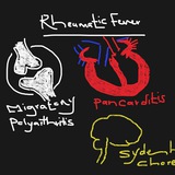
Rheumatic fever
Endocarditis
Myo , pericarditis
Valvular disease
Rheumatic fever
Endocarditis
Myo , pericarditis
Valvular disease
Q1. A 63-year-old man with small cell carcinoma of the left mainstem bronchus begins chemotherapy. During the treatment period, he becomes febrile and develops a productive cough. The temperature is 38.7°C respirations are 32 per minute, and blood pressure is 125/85 mm Hg. A CBC shows leukocytosis (WBC = 18,500/μL). The patient’s cough worsens, and he begins expectorating large amounts of foul smelling sputum. A chest X-ray shows a distinct cavity with an air/fluid level distal to the tumor area. Which of the following is the most likely diagnosis?
(A) Atelectasis
(B) Bronchiectasis
(C) Ghon complex
(D) Lobar pneumonia
(E) Pulmonary abscess
(A) Atelectasis
(B) Bronchiectasis
(C) Ghon complex
(D) Lobar pneumonia
(E) Pulmonary abscess
Q2.A 60-year-old alcoholic woman presents to the emergency room with fever, chills, and shortness of breath. The sputum is rusty-yellow and contains numerous neutrophils, red blood cells, and Gram-positive cocci. A chest X-ray shows diffuse haziness over both lungs. One week following admission, the patient develops empyema. This pulmonary condition is associated with the spread of bacterial infection to which of the following anatomic locations?
(A) Blood
(B) Bronchi
(C) Interstitial space
(D) Pericardium
(E) Pleural space
(A) Blood
(B) Bronchi
(C) Interstitial space
(D) Pericardium
(E) Pleural space